Tokyo, 2 May, /AJMEDIA/
Fujitsu and the Southern Tohoku General Hospital have launched a joint research project on an AI technology for the early detection of pancreatic cancer from computed tomography (CT) scans without contrast agent (non-contrast CT scans).
The new AI technology has been trained with data from 300 anonymized CT images of pancreatic cancer patients provided by the Southern Tohoku General Hospital and offers an optimal image analysis method based on the shape of organs and cancer tumors. The joint research project represents a new approach to apply AI technology to support medical practitioners in detecting early signs of pancreatic cancer from CT scans.
Fujitsu and the Southern Tohoku General Hospital will conduct clinical trials within this joint research to further improve the newly developed AI and offer a technology for an early detection of pancreatic cancer that can help to improve the quality of life of each patient.
The 5-year relative survival rate for people diagnosed with pancreatic cancer is 11% which is lower in comparison to other cancer types, as it is the fastest growing type of all cancers and generally difficult to detect. As the pancreas is located in the deepest part of the human body, affected people often don’t easily notice any symptoms and thus often don’t undergo voluntary medical examination until the cancer progresses. The fact that the entire pancreas is hard to visualize using simple imaging tests such as abdominal ultrasonography and that affected body parts are difficult to identify makes early detection of pancreatic cancer even more difficult.
Researchers anticipate that the development of technology capable of detecting microscopic signs of cancer from imaging data and improving test capabilities will play an important role in the early detection of pancreatic cancer.
The goal of this joint research is to successfully develop an AI technology that is able to detect signs of pancreatic cancer not only from contrast-enhanced CT scans (scans using contrast agents to make organs more visible) but also from non-contrast CT scans (scans not using contrast agents) to ultimately support the early diagnosis of affected patients regardless of symptoms of pancreatic cancer.
Outline of the joint research
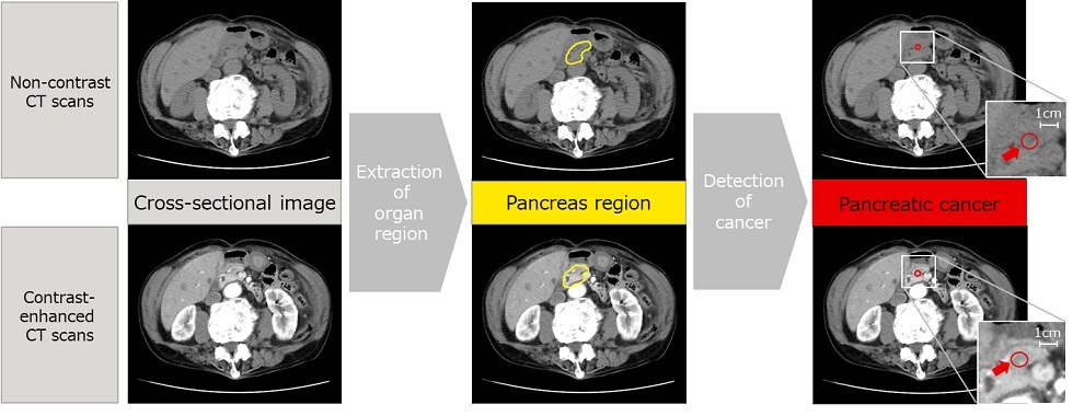
The precise and early detection of signs of pancreatic cancer in non-contrast CT scans represents a difficult task due to the low contrast of images and the unclear boundaries between the pancreas and other organs. To address this issue, Fujitsu and Southern Tohoku General Hospital aim to develop an AI technology that can identify the region corresponding to the pancreas (part marked yellow in figure 1) and detect the suspected parts affected by cancer (part marked red in figure 1). The technology achieves this by estimating the continuity between the anterior and posterior cross-sectional images in consideration of the anatomical tissue connection and automatically performs three-dimensional analysis including the anterior and posterior cross-sectional images in areas with strong continuity and planar analysis in areas with weak continuity.
The partners will also apply the newly developed technology in clinical practice to locate typical signs of early pancreatic cancer, including tumors and pancreatic duct dilatation that are often difficult to detect, as well as for findings that require clinical follow-up including cysts and local atrophy of the pancreas.